Айтон – раствор капель глазных на основе рецептов аюредической медицины. Обладает антисептическими свойствами, защищает глаза от загрязнений и вредного воздействия окружающей среды (пыль, дым, чрезмерная температура и пр.), успокаивает их. Нормализует нарушения зрительной функции, возникшие в результате заболеваний, помогает поддержанию остроты зрения при постоянном зрительном переутомлении.
Состав и форма выпуска
Айтон – раствор капель для глаз стерильный, содержит водные экстракты следующих компонентов: Azadirachta Indica 5%, Morinda pterygosperma 5%, Eclipta alba 5%, Boerhaavia diffusa 7%, Vitex nedundo 5%, Rosa damascena 7%, Carum corticum 2%, Terminalia chebula 5%, Terminalia belerica 2%, Emblica officinalis 5%, Curcuma longa, Camphora officinarum 3%, Mentha sylvestris 2%, Rock salt 2 %, Honey, Elettaria cardamomum, Santalum album, Pearl 1 %, Santalum rubrum 5%, Heliotropium indicum 5%, Ocimum sanctum 5%.
Форма выпуска: флаконы белые пластиковые с крышкой-дозатором по 10 мл в пачке из картона.
Фармакологические свойства
Раствор Айтон является тоником-эликсиром, созданным для гигиены глаз, а также поддержания высокой остроты зрения.
В его составе уникальная комбинация из лекарственных растений и определенных минералов, которая приготовлена в условиях современного фармацевтического производства по канонам традиционной аюрведической медицины.
При инстилляции раствор легко проникает ко внутренним структурам глаза и активирует циркуляцию внутриглазной жидкости, восстанавливает и гармонизирует функциональные системы органа зрения. Результатом подобного воздействия становится улучшение остроты зрения и здоровый блеск глаз.
Свойства компонентов раствора Айтон помогают защищать глаза от пыли, дыма и чрезмерного их пересушивания. Снимают усталость, раздражение и покраснение. Средство эффективно для поддержания функции зрения в случае, когда глаза подвергаются ежедневным высоким зрительным нагрузкам (работа на компьютере, с текстовыми документами и пр.).
В глазных каплях не заявлено вредных химических составляющих, они абсолютно натуральны. Их применение безболезненно и не раздражает глаз.
Показания к применению
Раствор Айтон эффективен для снятия напряжения зрения при длительной работе за компьютером, помогает скорейшей адаптации зрения при ослеплении светом (фары машин, отраженный свет в горах и пр.), кроме того, он обладает дезинфицирующими свойствами. Применяется при:
- Переутомлении глаз.
- Слезотечении.
- Снижении остроты зрения.
- Покраснении глаз.
- Аллергии.
- Ячмене.
- Сухости глаз.
- Рези в глазах.
Раствор также может применяться в комплексной терапии коньюктивитов, трахомы, блефаритов, изъязвлений роговицы, кератитов, помутнения хрусталика, глаукомы, близорукости, дальнозоркости и пр.
Способ применения и дозы
Стандартная дозировка раствора Айтон составляет 1-2 капли ежедневно конъюнктивально в начале и в конце дня. Однако, эта схема может быть изменена лечащим врачом. Частоту применения можно увеличить до 3-4 раз ежедневно с равными промежутками.
В случае конъюнктивита или иного инфекционного заболевания, раствор рекомендуется применяться до 6-ти раз в день с двухчасовым интервалом. В случае увеличения раздражения глаз, препарат необходимо отменить и проконсультироваться с врачом.
При катаракте и глаукоме, раствор может использоваться по стандартной схеме длительно – до 6-ти месяцев.
Противопоказания
Индивидуальная непереносимость какого-то из компонентов средства.
Побочные действия
Возникновение местной аллергической реакции.
Передозировка
Данных о случаях передозировки раствором Айтон нет.
Лекарственные взаимодействия
Не изучались.
Особые указания
Раствор капель для глаз Айтон является тоником для глаз не обладая терапевтическими свойствами.
Не содержит консервантов, поэтому при закапывании средства необходимо избегать соприкосновения дозатора с любыми поверхностями.
Для хранения раствора Айтом стоит выбирать удаленные от света места с комнатной температурой.
Срок годности продукта – 3 года. Однако, после вскрытия флакона, раствор годен не более месяца.
Цена препарата Айтон
Стоимость препарата «Айтон глазные капли» в аптеках Москвы начинается от 172 руб.
Аналоги Айтон

Артелак |

Дефислез |

Оксиал |

Хило-Комод |
Обратившись в «Московскую Глазную Клинику», Вы сможете пройти обследование на самом современном диагностическом оборудовании, а по его результатам – получить индивидуальные рекомендации ведущих специалистов по лечению выявленных патологий.
Мы работаем для Вас семь дней в неделю без выходных, с 9:00 до 21:00. Телефон для справок и записи на прием 8 (499) 322-36-36.
Также Вы можете воспользоваться формами ОНЛАЙН КОНСУЛЬТАЦИИ ВРАЧА и ОНЛАЙН ЗАПИСЬЮ на сайте.

Запишитесь на прием к врачу офтальмологу
Заполните форму и получите скидку 15 % на диагностику!
Глазные капли Айтон — лечебно-профилактическое средство и эликсир для глаз. Помогает сохранить остроту зрения и полезны при лечении различных глазных заболеваний. Капли действуют мягко и не вызывают жжения.
Глазные капли Айтон помогают нормализовать нарушения глазной функции, возникающие в результате различных заболеваний, таким образом, усиливая процесс излечения. Капли также помогают улучшению и поддержанию остроты зрения при любой работе, при которой идет постоянная нагрузка на глаза.
Показания к применению:
Показаниями к применению препарата Айтон являются:
— глаукома и катаракта, помутнение хрусталика глаза ;
— глазное давление;
— конъюнктивиты и пр. воспалительные процессы ;
— глаукома и катаракта помутнение хрусталика глаза ;
— снижение остроты зрения и утомление глаз ;
— слезоточивость ;
— эпифора, ретенционное слезотечение ;
— постоянная нагрузка на зрение и защита от неблагоприятных условий типа дыма сухости и жара ;
— покраснение и резь в глазах, головная боль от длительного просмотра телевизора или работы за компьютером ;
— кровоизлияния в стекловидное тело глаз.
Способ применения:
1 или 2 капли Айтон в каждый глаз вечером и после умывания утром (в соответствии с указанием врача).
Частота применения может быть увеличена до 3-4 раз в день с равными интервалами.
При конъюнктивите и других острых инфекциях глаз применять капли можно каждые два часа (по указанию врача).
При глаукоме, катаракте и иридоциклите может потребоваться курс лечения до 6 месяцев.
Условия хранения:
Капли Айтон следует хранить в сухом, темном и недоступном для детей месте.
Форма выпуска:
Айтон — глазные капли.
Флакон: 10 мл.
Состав:
Айтон содержит: стерильный раствор, содержащий водные экстракты следующих индийских растений: (in v/v)Nimba (Azadirachta Indica) 5%-Ниим Азадиратха Индийская, Sobhanjana (Morinda pterygosperma) 5%-Моринда, Bhringaraj (Eclipta alba) 5%- Эклипта Белая, Punarnava (Boerhaavia diffusa) 7,5%- Боэрхавия Размытая, Nirgundi (Vitex nedundo) 5%- Витекс, Satapatri (Rosa damascena) 7,5%-Роза Дамасская, Yamani (Carum corticum) 2 %-Карум, Haritaki (Terminalia chebula) 5%-Терминалия Хебула Миробалан Хебула, Vibhitaka (Terminalia belerica) 5%-Терминалия Белерическая Миробалан Белерический, Dhatriphala (Emblica officinalis)5%-Эмблика Лекарственная, Haridra (Curcuma longa) 5%- Куркума Длинная, Karpuram (Camphora officinarum) 3%-Камфора, Pudina (Mentha sylvestris) 2 %- Мята Болотная, Saindhava Laban (Rock salt) 2 %-Горная соль, Madhu (Honey) 10%-Мед, Ela (Elettaria cardamomum) 5%-Кардамон Элеттария, Swet Chandan (Santalum album) 5%-Белый Сандал, Mukta (Pearl) 1 %- кальцинированный жемчуг, Rakta Chandan (Santalum rubrum) 5%-Сандал, Hastisundi (Heliotropium indicum) 5% Гелиотроп Индийский, Tulsi Patra (Ocimum sanctum) 5%-лист Базилика Священного.
Аюрведические глазные капли «Айтон» — интересное сочетание традиционной индийской медицины и современных научных достижений. Ингредиенты, входящие в состав капель, эффективно используются в аюрведе для лечения глазных заболеваний и сохранения остроты зрения.
Глазные капли Aйтоун — абсолютно стерильный антисептический раствор, который успокаивает глаза, защищает их от различных загрязнений и вредных воздействий окружающей среды (дым, пыль, пары, чрезмерная температура и т.д.). Глазные капли AЙТОН помогают нормализовать нарушения глазной функции, возникающие в результате различных заболеваний, таким образом, усиливая процесс излечения. Капли также помогают улучшению и поддержанию остроты зрения при любой работе, при которой идет постоянная нагрузка на зрение.
«Айтон» в отличии от аюрведических капель «Уджала» и «Дришти» действуют более мягко и не вызывают жжения. Могут использоваться в течение дня. Ежедневно наше зрение подвергается стрессу. Работа за компьютером, просмотр телевизора, вредное воздействие окружающей среды, чтение в транспорте, несбалансированное питание. Всё это приводит к ослаблению зрения.
Своевременный уход за глазами предотвратит появление заболеваний и сохранит хорошее зрение в течение всей жизни.
Показания к применению
- профилактика глазных заболеваний
- воспаление глаз
- конюктивиты
- глазное давление
- снижение остроты зрения
- напряжение глаз
- переутомление
- глаукома и катаракта.
Способ применения: 1-2 капли в каждый глаз вечером и после умывания утром (в соответствии с указанием аюрведического врача). Частота применения может быть увеличена до 3-4 раз в день с равными интервалами. При конъюнктивите и других острых инфекциях глаз применять капли можно каждые два часа (по указанию врача). При глаукоме ,катаракте и иридоциклите может потребоваться курс лечения до 6 месяцев.
Если возникает раздражение и жжение в глазах, прекратите применение и проконсультируйтесь со своим аюрведическим доктором.
Большинство аюрведических капель вызывают лёгкое пощипывание или ощущение жжения глаз после закапывания. Это нормально и безопасно. После закапывания прилягте с закрытыми глазами на 5 минут, не протирая и не смывая капли. Так глаза смогут очиститься, и со слезами выйдут загрязнения. После этого жжение уйдёт, а ощущение в глазах станет приятным, лёгким и свежим!
Состав: Стерильный раствор, содержащий водные экстракты следующих индийских растений: Nim (Azadirachta Indica) 5%-Ним (Азадиратха Индийская), Sobhanjana (Morinda pterygosperma) 5%-Моринда, Bhringaraj (Eclipta alba) 5%- Эклипта Белая, Punarnava (Boerhaavia diffusa) 7,5%- Боэрхавия Раскидистая, Nirgundi (Vitex nedundo) 5%- Витекс, Satapatri (Rosa damascena) 7,5%-Роза Дамасская, Yamani (Carum corticum) 2 %-Карум, Haritaki (Terminalia chebula) 5%-Терминалия Хебула Миробалан Хебула, Vibhitaka (Terminalia belerica) 5%-Терминалия Белерическая Миробалан Белерический, Dhatriphala (Emblica officinalis)5%-Эмблика Лекарственная, Haridra (Curcuma longa) 5%- Куркума Длинная, Karpuram (Camphora officinarum) 3%-Камфора, Pudina (Mentha sylvestris) 2 %- Мята Болотная, Saindhava Laban (Rock salt) 2 %-Горная соль, Madhu (Honey) 10%-Мед, Ela (Elettaria cardamomum) 5%-Кардамон Элеттария, Swet Chandan (Santalum album) 5%-Белый Сандал, Mukta (Pearl) 1 %- кальцинированный жемчуг, Rakta Chandan (Santalum rubrum) 5%-Сандал, Hastisundi (Heliotropium indicum) 5% Гелиотроп Индийский, Tulsi Patra (Ocimum sanctum) 5%-лист Базилика Священного.
Объём: 10мл
ITONE (айтон) — аюрведические глазные капли, настоящий эликсир для глаз
Глазные капли Айтон (Itone Eye Drops) – аюрведические капли с экстрактами целебных трав, для профилактики и лечения глазных заболеваний и повышения остроты зрения.
Глазные капли Айтон оказывают успокаивающее и защитное действие на глаза: снимают глазное напряжение, расслабляют и защищают от внешнего воздействия пыли, ветра, микробов и от вредного излучения при работе за компьютером. Это средство помогает восстановить правильную работу органов зрения, нарушенную по причине каких-либо заболеваний. Капли поддерживают и повышают остроту зрения.
Эти капли оказывают мягкое воздействие на слизистую, не вызывают раздражение и зуд. Их можно использовать на протяжении всего дня. Айтон способствует профилактике глазных заболеваний и нарушений зрения, которые так часто возникают у людей любого возраста в современном мире. Капли снимают стресс и глазное напряжение при многочасовой работе за компьютером.
Также они эффективны при лечении серьёзных заболеваний, таких как катаракта и глаукома.
Регулярный уход за глазами поможет избежать глазных заболеваний и сохранить прекрасное зрение на всю жизнь.
Полезные свойства
- Снимает воспаление, покраснение, раздражение глаз;
- Повышает остроту зрения;
- Профилактика глазных заболеваний;
- Защита от внешних факторов (пыль, ветер, солнце и т. д.);
- Снимает напряжение и усталость глаз;
- Нормализует глазное давление;
- Восстановление зрения.
Показания к применению
- Глаукома и катаракта,
- Глазное напряжение,
- Конъюнктивит,
- Воспалительные процессы глаз,
- Ухудшение зрения,
- Покраснение и резь в глазах
- Слезоточивость,
- Работа за компьютером,
- Защита от неблагоприятных условий (дым, пыль, пар, сухость, жар).
Способ применения
Применять утром и вечером после умывания по 1 – 2 капли.
При коньюктивите можно закапывать каждые 3 часа.
При лечении глаукомы и катаракты необходим длительный курс от 6 до 12 месяцев, по рекомендации врача.
Состав
Cтерильный раствор, с содержанием водных экстрактов индийских растений: (in v/v) Nimba (Azadirachta Indica) 5% – Ниим Азадиратха Индийская Sobhanjana (Morinda pterygosperma) 5% – Моринда Bhringaraj (Eclipta alba) 5% – Эклипта Белая Punarnava (Boerhaavia diffusa) 7,5% – Боэрхавия Размытая Nirgundi (Vitex nedundo) 5% – Витекс Satapatri (Rosa damascena) 7,5% – Роза Дамаская Yamani (Carum corticum) 2 % – Карум Haritaki (Terminalia chebula) 5% – Терминалия Хебула Миробалан Хебула Vibhitaka (Terminalia belerica) 5% – Терминалия Белерическая Миробалан Белерический Dhatriphala (Emblica officinalis)5% – Эмблика Лекарственная Haridra (Curcuma longa) 5% – Куркума Длинная Karpuram (Camphora officinarum) 3% – Камфора Pudina (Mentha sylvestris) 2 % – Мята Болотная Saindhava Laban (Rock salt) 2 %-Горная соль Madhu (Honey) 10% – Мед Ela (Elettaria cardamomum) 5% – Кардамон Элеттария Swet Chandan (Santalum album) 5% – Белый Сандал Mukta (Pearl) 1 % – кальцинированный жемчуг Rakta Chandan (Santalum rubrum) 5% – Сандал Красный Hastisundi (Heliotropium indicum) 5% – Гелиотроп Индийский Tulsi Patra (Ocimum sanctum) 5% – лист Базилика Священного Консерванты: Benzalkonium Chloride 0,01 % w/v Phenylethyl alcohol 0,05% v/v.
Противопоказания
Индивидуальная непереносимость отдельных компонентов. При беременности и в период лактации необходимо проконсультироваться с врачом.
Все товары, представленные на сайте Himalay.ru в разделе «Средства аюрведы», не являются лекарствами, а относятся к фитосборам или добавкам к питанию. Перед применением рекомендуется проконсультироваться со специалистом по Аюрведе для индивидуального назначения.
Описание и применение Глазные капли Айтон (ITONE), 10 мл
Аюрведические глазные капли «Айтон» — интересное сочетание традиционной индийской медицины и современных научных достижений. Ингредиенты, входящие в состав капель, эффективно используются в аюрведе для лечения глазных заболеваний и сохранения остроты зрения.
Капли «Айтон» стерильны и безвредны, снимают усталость и напряжение глаз, успокаивают, очищают от мельчайших частичек пыли, убирают покраснение, снимают воспалительные процессы, коньюктивиты , также, с помощью этих капель, лечатся такие серьезные заболевания как глаукома и катаракта.
«Айтон» в отличии от аюрведических капель «Уджала» и «Дришти» действуют более мягко и не вызывают жжения. Могут использоваться в течение дня. Ежедневно наше зрение подвергается стрессу. Работа за компьютером, просмотр телевизора, вредное воздействие окружающей среды, чтение в транспорте, несбалансированное питание. Всё это приводит к ослаблению зрения.
Своевременный уход за глазами предотвратит появление заболеваний и сохранит хорошее зрение в течение всей жизни. Показания к применению: — профилактика глазных заболеваний; — воспаление глаз; — конюктивиты; — глазное давление; — снижение остроты зрения; — напряжение глаз, переутомление; — глаукома и катаракта.
Компоненты капель Айтон:
Cтерильный раствор, с содержанием водных экстрактов индийских растений: (in v/v)Nimba (Azadirachta Indica) 5% — Ниим Азадиратха Индийская Sobhanjana (Morinda pterygosperma) 5% — Моринда Bhringaraj (Eclipta alba) 5% — Эклипта Белая Punarnava (Boerhaavia diffusa) 7,5% — Боэрхавия Размытая Nirgundi (Vitex nedundo) 5% — Витекс Satapatri (Rosa damascena) 7,5% — Роза Дамаская Yamani (Carum corticum) 2 % — Карум Haritaki (Terminalia chebula) 5% — Терминалия Хебула Миробалан Хебула Vibhitaka (Terminalia belerica) 5% — Терминалия Белерическая Миробалан Белерический Dhatriphala (Emblica officinalis)5% — Эмблика Лекарственная Haridra (Curcuma longa) 5% — Куркума Длинная Karpuram (Camphora officinarum) 3% — Камфора Pudina (Mentha sylvestris) 2 % — Мята Болотная Saindhava Laban (Rock salt) 2 %-Горная соль Madhu (Honey) 10% — Мед Ela (Elettaria cardamomum) 5% — Кардамон Элеттария Swet Chandan (Santalum album) 5% — Белый Сандал Mukta (Pearl) 1 % — кальцинированный жемчуг Rakta Chandan (Santalum rubrum) 5% — Сандал Красный Hastisundi (Heliotropium indicum) 5% — Гелиотроп Индийский Tulsi Patra (Ocimum sanctum) 5% — лист Базилика Священного Консерванты:Benzalkonium Chloride 0,01 % w/v Phenylethyl alcohol 0,05% v/v
Способ применения: применять утром и вечером после водных процедур по 1-2 капли. При коньюктивите можно закапывать каждые 3 часа. При лечение глаукомы и катаракты необходим длительный курс от шести месяцев до года, по рекомендации врача. Хранить в сухом прохладном месте.
БАД не является лекарством. Необходима консультация специалиста.
Отзывы о Глазные капли Айтон (ITONE), 10 мл
Отличные капли, утром и вечером закапываю, главное проморгаться как следует, лечебное ощущение. Спасибо, всех благ!
Добрый день. Получила заказ 59824. Капельки отлично снимают раздражение , усталость и сухость после рабоы за компьютером. Пользуюсь уже несколько месяцев. Магазин очень нравиться.
[size=100]Заказ [/size]#55937 получили. Капли заказываю второй раз в этом же магазине, так как результат был отличным. С глаз вроде пелена ушла. Стала видеть очень мелкий шрифт. Да и в глазах ощущение свежести. Замечательные капли. Всем рекомендую.
Это невероятно, но факт! Я тоже стал лучше разбирать мелкий шрифт. «Мушки» в глазах также уменьшились.
Хорошие капельки. Отлично снимают усталость глаз и не щипят.
Эти чудесные капли для глаз заказываю в третий раз. Мало того,теперь ими пользуются 4 моих коллеги. Эффект после закапывания моментальный и прекрасный: глаза успокаиваются, уходит «пелена» перед глазами, уменьшается сухость конъюктивы, полученной вследствие долгой работы за компьютером. Заказ последний 43456. Обязательно ещё закажу и друзьям посоветую.
Капли работают. Через примерно месяц использования улучшилось близкое зрение, стала лучше разбирать мелкий шрифт.
Получил заказ 30834 пробовал разные капли эти в первый раз начал капать
капнул один раз понравились нет жжения очень мягкие что будет дальше отпишу понравилось
работа компании быстро , качественно , собираюсь заказывать постоянно
Спасибо
Эти капельки купила впервые, но так как очень доверяю аювердическим средствам, да и при моих проблемах со зрением не навредить ,это главное. Спасибо! Понравились!
Впервые попробовала эти капли и совершенно не пожалела. Теперь с ними не расстаюсь.
Практически моментально снимает усталось и неприятные ощущения.
Даже складывается ощущение, что после использования на какое то время лучше вижу.
Первые дня три немного жгло глаза первые секунд пять, потом прекратилось.
Заказываю эти капли постоянно, в восторге от их эффективности. Пробывала много аптечных средств для глаз, и всеми была разочарована, уверилась что только капли индийского производства на самом деле помогают!
Такие капли покупали год назад в Индии для жены — у нее сухость и слезотечение были
вечером после работы, ничего не помогало, испробовали десятки видов капель. Начала капать эти капли
и через некоторое время (примерно пару недель) эффект оказался великолепным — слезоточивость прошла белки посветлели,приятное чувство чистоты.
Теперь нашли их в этом интернет магазине, будем заказывать постоянно. Посоветую эти капли всем кто страдает от сухости и песка в глазах.
заказ #18751 привезла изначально маме эти капли из Индии, ей понравились, сказала — лучшие капли для глаз, что она пробовала, надолго и быстро снимают усталость, сама я пробовала — жжется, но помогает, отличные капли, маме беру постоянно
[size=100]Одни из самых мягких капель по ощущениям после закапывания. Я бы сказала, что практически совсем не щиплют. Кто знаком с индийскими каплями, тот знает, что многие из них очень сильно жгут в первые несколько секунд после закапывания. Эти капли вполне нейтральные, можно закапывать даже в течение дня, слезотечения не вызывают. Состав хороший, может быть не такой лечебный, как, например, у Уджалы, но с функцией снятия напряжения, усталости и устранения сухости справляются отлично. Я ношу контактные линзы, капли использую только после снятия линз. Заказ № 17860.[/size]
Каплями пользуясь четвёртый год. Я ношу линзы, после снятия линзы остаётся ощущение сухости, капли хорошо увлажняют, снимают раздражение. Приобретают капли не только для себя, но уже для шести моих коллег, все очень довольны.
Пришло быстро, описанию соответствуют, не щипят, понравились, рекомендую
Хорошие. Понравились. Пока это — самые мало щиплющие капли из всех традиционных индийских. И состав хороший.
Отличные капли для снятия усталости и сухости глаз. Капли не жгут, поэтому можно закапывать в любое время, когда это необходимо. Для людей, проводящих много времени за компьютером, отличное средство. Эффект — отдохнувшие, выспавшиеся, блестящие глазки. Очень довольна покупкой.
- Ним (Azadirachta indica).
- Трифала (Triphala).
- Амла (Emblica officinalis).
- Пунарнава (Boerhavia diffusa).
- Гималайская розовая соль (Saindhava lavana).
- Харитаки (Terminalia chebula).
- Мед (Honey).
- Роза столистная (Rosa centifolia).
- Коричник камфорный (Cinnamomum camphora).
- Туласи (Ocimum sanctum).
Растение, которое пользуется широкой известностью как в Индии, так и за ее пределами. Благодаря натуральным антибактериальным свойствам эффективно борется с микробами и раздражением глаз, снимает зуд. Оказывает выраженный антисептический эффект.
Лечебный состав из трех целебных трав: амла, харитаки, бибхитаки. Относится к Расаянам — средствам для омолаживания всех тканей организма. Выводит токсины и шлаки. Богат витаминами и минералами, в особенности витамином C. Способствует улучшению зрения.
Применяется также для улучшения зрения, предотвращения возникновения возрастных глазных заболеваний, таких как глаукома, катаракта, пресбиопия, способствует снижению внутриглазного давления, расслабляюще и успокаивающе действует на мышцы и оболочки глазного яблока.
Одно из самых почитаемых в Аюрведе растений, на санскрите «пунарнава» буквально означает «снова новый». Имеет мощный антиоксидантный и противовоспалительный эффект. Снижает болезненные ощущения в области глаз, уменьшает слезоотделение, улучшает обменные процессы и клеточное питание. Замедляет развитие катаракты. Обладает вяжущими, кровоостанавливающими и заживляющими свойствами.
Содержащиеся в гималайской соли 84 полезных для организма микроэлемента улучшают обменные процессы в организме. Она имеет ярко выраженные антигистаминные и антибактериальные свойства, помогает при воспалительных заболеваниях, помогает избавиться от аллергических реакций, угнетает рост болезнетворной микрофлоры, защищает от грибков, микробов и вирусов.
Известное омолаживающее растение. Улучшает зрение. Противовоспалительное, антибактериальное средство.
Хорошее противовоспалительное средство для глаз, которое к тому же содержит огромное количество витаминов и микроэлементов. Устраняет боль в глазах и их усталость, нормализует высокое глазное давление, снимает воспаление век, улучшает зрение.
Известна своими охлаждающими свойствами. Быстро снимает напряжение глаз и помогает при компьютерном зрительном синдроме (астенопии). Имеет антибактериальные свойства.
Имеет охлаждающий эффект, предохраняет глаза от инфекций, смягчает кожу вокруг глаз, укрепляет ресницы и ускоряет их рост.
Защищает глаза от различных заболеваний, таких как глаукома и дегенерация желтого пятна. Она также успокаивает глаза и снижает нагрузку, вызванную постоянным использованием компьютера.
Основная информация:
- содержит натуральные компоненты
- улучшает состояние при глазных заболеваниях
- устраняет нарушения функций глаз, появившиеся как результат различных заболеваний
- поддерживает остроту зрения при длительной работе за компьютером или продолжительном чтении
- улучшает состояние при конъюнктивите
- снимает напряжение и усталость глаз
- устраняет покраснения и сухость
- не является лекарственным средством
Показания к применению:
- глазные заболевания
- снижение остроты зрения
- защита глаз
- конъюнктивит
- напряжение
- усталость глаз
- покраснение глаз
- сухость глаз
Противопоказания:
- индивидуальная непереносимость отдельных компонентов
Активнодействующие компоненты:
Амла (E. officinalis) – предотвращает появление глаукомы, катаракты, снижает внутриглазное давление, улучшает зрение.
Ним (A. indica) – снимает раздражения и зуд, оказывает противовоспалительное и антисептическое действия.
Моринга масличная (M. Pterygosperma) – устраняет боль, снимает усталость, нормализует внутриглазное давление.
Роза столистная (R. centifolia) – защищает от инфекций, уменьшает слезоотделение, предотвращает развитие катаракты.
Инструкция по применению:
Для профилактики снижения остроты зрения, при красноте, постоянном напряжении глаз – капать 2 раза в день по 1-2 капли.
При конъюнктивите и прочих острых инфекциях – капать каждые 2-3 часа по 1-2 капли.
Рекомендуется предварительная консультация с врачом.
Состав:
Каждые 10 мл содержат:
Водные дистилляты: Ним (A. indica) – 562.50 мг, Моринга масличная (M. Pterygosperma) – 562.50 мг, Брингарадж (E. alba) – 562.50 мг, Пунарнава (B. diffusa) – 562.50 мг, Прутняк китайский (V. negundo) – 562.50 мг, Тулси (O. sanctum) – 562.50 мг, Роза столистная (R. centifolia) – 150 мг, Ажгон (C. copticum) – 69.50 г, Харитаки (T. chebula) – 46.25 мг, Бибхитаки (T. belerica) – 46.25 г, Амла (E. officinalis) – 46.25 мг, Куркума длинная (C. longa) – 92.50 мг, Камфора (Camphor) – 6.95 мг, Ментол (Menthol) – 1.85 мг, Мед (Honey) – 92.50 мг, Черный кардамон (A. subulatum) – 27.75 мг, Сандал белый (S. album) – 69.50 мг, Жемчужный кальций (Pearl) – 0.93 мг, Каменная соль (Rock salt) – 13.88 мг.
Консерванты: Фенилэтиловый спирт (Phenethyl alcohol) – 0.05%, Бензалкония хлорид (Benzalkonium chloride) – 0.02%.
Условия хранения:
Хранить в сухом недоступном для света месте.
Отказ от ответственности:
MYINDIA.RU старается предоставлять только актуальную информацию о продукции, но дизайн упаковки может отличаться от представленного на сайте. Перед использованием препарата ознакомьтесь с приложенной к нему инструкцией по применению или проконсультируйтесь со специалистом.
Вес: 45 г.
- Research article
- Open Access
- Published: 02 January 2013
- Pankaj Gupta1,
- Alok Kumar Ravi1,
- Hanuman Prasad Sharma1 &
- …
- Nihar Ranjan Biswas2
BMC Complementary and Alternative Medicine
volume 13, Article number: 1 (2013)
Cite this article
-
9114 Accesses
-
97 Citations
-
2 Altmetric
-
Metrics details
Abstract
Background
The polyherbal eye drop (Itone™) is a mixture of aqueous distillates of nineteen traditionally used ingredients that sum up to impart potency to the formulation and make it a useful adjunct in various ocular pathologies. However, as there have been no controlled experimental studies accounting to the above claim, therefore, the present study was designed to evaluate the polyherbal formulation (PHF) for antiangiogenic, anti-inflammatory, anticataract, antioxidant and cytotoxicity in addition to the evaluation of intraocular penetration of PHF in rabbit eyes using LC-MS/MS.
Materials and methods
Antiangiogenic activity of the PHF was evaluated using in ovo chick chorio-allantoic membrane (CAM) assay and in vivo cautery induced corneal neovascularization assay in rats. Anticataract potential was evaluated using steroid induced cataract in developing chick embryos, sodium selenite induced cataract in rat pups and galactose induced cataract in rats. The antioxidant activity was evaluated using di-phenyl picryl hydrazyl (DPPH) radical scavenging assay. Anti-inflammatory activity was evaluated in vitro using inhibition of LTB4 formation in human WBCs and in vivo using carrageenan induced paw edema assay in rats. The cytotoxicity was evaluated against HeLa cancer cell lines using (3-(4,5-Dimethylthiazol-2-yl)-2,5-diphenyltetrazolium bromide (MTT) assay. Furthermore evaluation of the intraocular penetration of the PHF was carried out in rabbit eyes via aqueous humor paracentesis and further analysis using LC-MS/MS.
Results
PHF significantly inhibited VEGF induced proliferation of new blood vessels in CAM assay and inhibited the cautery induced corneal neovascularization in rats. Additionally, PHF showed noticeable delay in the progression of cataract in the selenite and galactose induced cataract models whereby the PHF treated lenses were graded for stages II and III respectively. However, the PHF did not show any anticataract activity in the hydrocortisone induced cataract model. Moreover, PHF exhibited anti-inflammatory activity whereby it showed 39.34% inhibition of LTB4 formation and significantly inhibited carrageenan induced paw edema in rats. Eight compounds of PHF viz. camphor, casticin, curcumin-II, quercetin, rosmarinic acid, γ-terpinene, β-pinene and dipentene exhibited transcorneal penetration in rabbit eyes.
Conclusion
The significant antiangiogenic and anti-inflammatory activities evinced by the PHF merits further investigation for ocular neovascular and inflammatory diseases in humans.
Peer Review reports
Background
The extracts of herbal products and Ayurvedic, Siddha and Unani formulations are mixtures of at least partially uncharacterized constituents. It is claimed that such a mixture provides a therapeutic advantage, since the unknown constituents may be additive or synergistic in action or may produce a balance to counteract adverse effects of any one constituent. This may provide more efficacy than the known constituent used alone. Thus, purification of the medicines is not required as it may lead to loss of the advantages provided by the mixture [1]. An ayurvedic proprietary polyherbal eye drop (Itone™) developed on the basis of ayurvedic principles have been available for the last 25 years in Indian Market and has been recognized to have medicinal value for various ocular ailments. The polyherbal formulation (Itone™) consists of a fixed combination of the aqueous distillates of nineteen traditionally used ingredients including Azadirachta indica (nimba-5%), Moringa pterygosperma (sobhanjana-5%), Eclipta alba (bhringaraj-5%), Boerhaavia diffusa (punarnava-7.5%), Carum copticum (yamani-2%), Terminalia chebula (haritaki-5%), Terminalia belerica (vibhitaka-5%), Emblica officinalis (dhatriphala-5%), Santalum album (swet chandan-5%), mukta (pearl-1%), Ocimum sanctum (tulsi patra-5%), Vitex negundo (nirgundi-5%), Curcuma longa (haridra-5%), Mentha piperata (menthol-2%), Cinnamomum camphora (camphor-3%), Amomum subulatum (ela-5%), Rosa centifolia (satapatri-7.5%), saindhava laban (rock salt-2%) and madhu (honey-10%). It has been reported to be a useful adjunct in various ocular diseases like conjunctivitis, trachoma, blepharitis, keratitis, corneal ulcers etc. It has been reported to maintain the visual acuity and helpful in computer vision syndrome [2, 3].
Several clinical studies are available appreciating the effect of Itone™ in various ocular pathologies. However, controlled experimental studies are not available to substantiate the claim. Therefore, the present study was carried out to systematically evaluate the above polyherbal formulation (PHF) for several ocular indications using contemporary technologies and experimental models to explore antiangiogenic, anticataract, anti-inflammatory, cytotoxicity and antioxidant properties. The above PHF was also subjected for the evaluation of its intraocular penetration after topical application in rabbits. These approaches are expected to increase the modern application of traditional knowledge for their scientific rationality and therapeutic application.
Methods
Chemical and reagents
Vascular Endothelial Growth Factor (VEGF) and MTT were purchased from Sigma-Aldrich, USA. Dulbecco’s Modified Eagle Medium (DMEM) was purchased from Invitrogen, India. Hydrocortisone sodium succinate injection was purchased from Alkem laboratories Ltd., Mumbai, India. Sodium selenite was purchased from Loba chemie, India. Riboflavin was purchased from Sisco Research Laboratories Pvt. Ltd., Mumbai, India. Galactose was purchased from Spectrochem Pvt. Ltd., Mumbai, India. The preservative free PHF (Itone™ eye drops, Batch No — 2142) were provided by Dey’s Medical Stores, Kolkata, India. All the other reagents were of analytical grade and were used without further purification.
All study protocols were approved by standing Institutional Animal Ethics Committee (IAEC) of All India Institute of Medical Sciences (AIIMS), New Delhi, India. All animal experiments were done in accordance with guidelines of Association for Research in Vision and Ophthalmology (ARVO).
Evaluation of antiangiogenic activity of the PHF
Chick Chorio-Allantoic Membrane (CAM) assay
The CAM assay was performed as per the procedure described by Ribatti et al., 1997 [4] with slight modifications. Briefly, fresh fertile white leghorn chick eggs were purchased from Kegg Farm (Gurgaon, Haryana, India) and further placed in a humidified incubator at 36 ± 2°C. On the third day of incubation, a hole was drilled on the egg shell and 2–3 ml of albumin was withdrawn aseptically. The hole was sealed using sterile parafilm and eggs were re-subjected for incubation. On seventh day, a suitable window was opened and VEGF (50 ng) in ovalbumin coated coverslips of 12 mm diameter (with or without test substance) were placed over the chorio-allantoic membrane. Coverslips coated only with ovalbumin, ovalbumin with VEGF, ovalbumin with VEGF and thalidomide (10 μg), ovalbumin with VEGF and PHF (50 μl) served as normal, control, positive control and test respectively. A minimum of 8 eggs were used per group. The windows were further sealed with sterile parafilm and eggs were reincubated for further period up till twelfth day. On the day 12, the blood vessels grown under the coverslip area were digitally photographed using a sensitive CCD camera directly attached to the computer using graphic user interface. The blood vessel areas were analysed using Aphelion Developer Image Analysis Software (Adcis, France).
Corneal neovascularization assay
Corneal neovascularization was induced by chemical cauterization, using the technique described by Mahoney & Waterbury,1985 [5]. One cornea of each rat was cauterized by pressing an applicator stick (with a diameter of 1.5 mm) coated with 75% silver nitrate / 25% potassium nitrate on the centre of the cornea for 5 sec while the animal was deeply anesthetized with sodium pentobarbitone. To increase the reproducibility of the injuries, the same investigator cauterized all animals. Following cauterization, the rats were randomized to eliminate potential bias in the degree of injury within the different groups. Rats were randomly divided into three groups (n = 6) viz. sham, positive control (bevacizumab 1.25 mg/ml) and PHF treated. 20 μl of test was applied topically to each cauterized cornea three times per day for 5 days starting from the day of cauterization. The first treatment with each medication was started approximately 30 min after cauterization. The corneal neovascularization was assessed on day 6 after cauterization whereby the corneas of all rats were subjected for photography using CCD camera. The photographs were further subjected for quantification of corneal neovascularization using the Aphelion Dev. image analysis software (Adcis, France).
Evaluation of PHF for its cytotoxicity
MTT assay
The HeLa cancer cell lines (No. 25) were obtained from National Centre for Cell Science (NCCS), Pune, India and suspended in 2.0 ml of DMEM (10% Fetal Bovine Serum). Cell count was adjusted to 5 × 10⁴ cells/ml. 100 μl of this cell suspension was plated in each well of 96-well ELISA plate. Plated cells were incubated in a CO2 incubator (5% CO2, 37°C) for 16 h, after which the media was removed from wells and fresh DMEM media containing 50 μl PHF was added in triplicates followed by further incubation for 24 h. After 24 h, DMEM media was removed and 100 μl of MTT (1 mg/ml) was added to each well followed by incubation for 3 h in CO2 incubator. After 3 h, media containing MTT was removed and 100 μl of DMSO was added to each well to dissolve the formazan crystals. Absorbance was taken at 570 nm and 655 nm.
Evaluation of anticataract potential of PHF
Hydrocortisone induced cataract in developing chick embryos
The procedure previously standardized in our laboratory [6] was used for this experiment. Briefly, ten days old White leghorn chick eggs were procured from Kegg Farm (Gurgaon, Haryana, India). The eggs were randomly divided into three groups (n =  namely normal (saline treated), control and PHF treated respectively. All the eggs were incubated in a humidified incubator at 36 ± 2°C. On 15th day, a hole was drilled in the eggshell over the air sac. Control and test groups received hydrocortisone (0.25 μM/0.1 ml) injected through the hole to induce cataract where as normal group received 0.1 ml of normal saline. The puncture was sealed with an adhesive tape and eggs were re-incubated. Control and test groups received 0.1 ml of normal saline and PHF at 3, 10 and 20 h after hydrocortisone injection respectively. After 48 h of the hydrocortisone injection the groups were sacrificed and the eyes were enucleated. The lenses were dissected out and were subjected for grading by two independent observers who were not aware of the treatment.
For the evaluation of lens, original grading of Nishigori et al., 1982 [7] was used, stage 1 — lens is clear and indistinguishable from normal (no cataract), stage 2 — faint opaque ring between the cortical region and nuclear region, stage 3 — distinct opaque ring between both the regions, stage 4 — lens has a pin hole size of clear area in opaque nucleus and stage 5 — opaque nucleus.
Selenite induced cataract model
Wistar female albino rats along with pups (6 days postnatal) were housed together. Nursing mother was fed with normal diet and water ad libitum. Two groups having mother and pups were divided. The pups of each rat were divided into two groups namely control and PHF treated and each group consisted of 6 pups respectively. The rat pups of the control and treated group were given sodium selenite at the dose of 25 μM/kg subcutaneously on the 9th postnatal day. Treated group received PHF subcutaneously in the volume of 0.05 ml from the seventh postnatal day to sixteenth day. The eyes were subjected for torchlight and slit lamp examination on the sixteenth postnatal day and compared with control for the presence and absence of nuclear cataract [8].
In vivo galactose induced cataract in rats
Wistar albino rats of either sex weighing 80–100 g were fed with 30% galactose diet with water ad libitum for 28 days. The rats were divided equally into two groups of control and test, each consisting of 5 animals. Test group was instilled with PHF amounting to 20 μl three times a day and control animals received saline. Each eye was evaluated for the presence of cataract and was graded by Sippel’s method [9] using slit lamp on 7, 14, 21 and 28th day.
Grading of lenses
Two independent observers those who were not aware of the treatment evaluated the stages of cataract using slit lamp biomicroscopy in rat eyes and mean of their grading was considered for further comparisons. The lenses were graded using the Sipple’s scale, Stage 1 — Clear lens, Stage 2 — Peripheral vacuoles, Stage 3 — Irregular peripheral vacuoles with the involvement of lens cortex, Stage 4 — Irregular opacity of lens and Stage 5 — Pronounced opacity.
Evaluation of the PHF for its antioxidant activity
DPPH radical scavenging assay
The free radical scavenging activity of PHF was measured in term of hydrogen donating or radical scavenging ability using the stable radical DPPH [10]. Briefly, 1.0 ml of 0.1 mM solution of DPPH in ethanol was added to 3.0 ml of test solution (with varying dilutions) and 30 min later, the absorbance was measured at 517 nm. Sodium ascorbate was used as a positive control and deionized water served as control. Percentage radical scavenging activity (%RSA) was calculated by following equation:
%RSA
=
Ab
s
control
−
Ab
s
test
/
Ab
s
control
*
100
Abscontrol is the absorbance of control in presence of deionized water without test and Abstest is the absorbance in presence of PHF or Sodium ascorbate. The antioxidant activity was expressed as IC50 (concentration (μg/ml) of the test that inhibited the formation of DPPH radicals by 50%).
Evaluation of PHF for its anti-inflammatory activity
Carrageenan induced paw edema
Overnight fasted Wistar albino rats (n = 6) of either sex (100–150 g) were divided into control (saline treated), positive control (diclofenac 5 mg/kg bodyweight treated) and experimental (4 ml/kg PHF treated) groups. After one hour of intraperitoneal drug administration, paw edema was induced by single injection of 0.1 ml of 1% carrageenan injected intradermally on the plantar side of the right hind paw. Paw volumes were measured using a digital plethysmometer (Inco, Haryana) immediately before the injection of carrageenan and at hourly intervals for 4 h thereafter. The volume of edema was expressed for each rat as the difference before and after the injection of carrageenan. The percent inhibition of paw edema was calculated by following equation:
%Inhibition
=
V
c
–
V
t
/
V
c
*
100
Vc is the paw volume of control that received saline and Vt is paw volume of test that received PHF or Diclofenac.
LeukotrieneB4 (LTB4) inhibition assay
Fresh buffy coat was obtained from hospital blood bank and subjected for WBC separation using HiSep (Hi-media, Mumbai) and diluted with equal volume of Hank’s balanced salt solution. The metabolism of endogenously bound arachidonic acid to LTB4 was measured in a total volume of 0.4 ml of the cell suspension at 37°C. The reaction tubes were containing 20 μg arachidonic acid and the investigational drugs (PHF and zileuton). After 45 min pre-incubation the reaction was started by addition of 20 μg of calcium ionophore and 2 μg of glutathione. After 15 min the reaction was stopped with 40 μl of 0.1 M HCl. After centrifugation for 2 min aliquots of the supernatant were subjected to LC-MS/MS estimation of LTB4.
Hypersil GOLD column (50 mm × 2.1 mm, 1.9 μm, Thermo, Waltham, MA, USA) with a gradient system of acetonitrile with 0.1% formic acid and water with 0.1% formic acid was used for chromatographic separation. The gradient was started with 80:20 water/acetonitrile and reached to 30:70 in 3 min and first line condition was achieved over a period of 2 min. Flow rate was maintained at 0.5 ml/min.
Tandem Mass spectrometric detection of analytes and internal standard (IS) was carried out with an electrospray ionization (ESI) source operated in the negative mode. Multiple Reaction Monitoring (MRM) mode was performed for quantification. 335.2 m/z was selected as precursor ion (Q1) whereas 317.2 m/z and 195.1 m/z (Q3) were selected as product ion for LTB4. Optimized compound dependent parameters were Declustering Potential (DP) — 76, Entrance Potential (EP) — 10, Collision Energy (CE) — 20 and Collision Cell Exit Potential (CXP) — 8. Probenecid was selected as an IS with 283.91 m/z (Q1) and 240 m/z (Q3).
Evaluation of intraocular penetration of PHF
New Zealand Albino Rabbits (n = 4) of either sex weighing 2–2.5 kg body weight were used for this study. Briefly 50 μl of PHF was instilled after each 10 min for 1 h into the cul-de-sac of the rabbit eyes. After completion of dosing, eyes were thoroughly washed with normal saline and aqueous humor was aspirated at 2 h via paracentesis. Briefly, eyes were anaesthetized by instilling 4% lidocaine and 70–100 μl of aqueous humor was withdrawn with the help of 30 G sterile hypodermic needle. All the samples were stored at −70°C until analysis by LC-MS/MS.
For the detection of compounds in PHF, information dependent acquisition (IDA) method using a hybrid system of Ultra High Performance Liquid Chromatography (UHPLC) coupled with Mass Spectrometry (LC-MS/MS) was used.
UHPLC (Accela, Thermo Surveyor system, Thermo Electron Corp, Waltham, MA, USA) was equipped with a quaternary pump connected to an online degasser and photodiode array (PDA) coupled with triple quadrupole mass spectrometer (4000 Q-Trap, AB/MDS Sciex, Foster City, CA, USA). Luna® column (250 mm × 4.6 mm, 5 μm, Phenomenex, USA) with a linear gradient system of water with 0.1% formic acid and acetonitrile with 0.1% formic acid was used for chromatographic separation. Gradient was started with 10% organic mobile phase and reached to 100% with 18% increase in each 5 min.
Tandem Mass spectrometric detection was carried out on an Applied Bio Systems 4000 triple quadrapole instrument (ABS Biosystems, Foster City CA, USA) equipped with an ESI source that operated in the positive ion mode. For a mass range from 100 to 800 atomic mass unit (amu), IDA scanning was done at a speed of 1000 Da/s with Enhanced Mass Scan (EMS). Chromatogram for PHF obtained from IDA scan was screened for the masses available in literature.
The analytes which were found to be present in IDA scan for PHF were further confirmed by observing their fragmentation patterns using MRM mode in LC-MS/MS. For MRM, all compounds were optimized for their fragmentation patterns and compound dependent parameters and subsequent separation of compounds was achieved using Purospher® STAR RP-18 (55 mm × 4 mm, 3 μm, Merck, Darmstadt, Germany) column with guard. For chromatographic separation a linear gradient of water with 0.1% formic acid and acetonitrile with 0.1% formic acid was used in a starting ratio of 90% acetonitrile. The gradient reached to 100% organic phase with an increment of 1% in 1 min.
The aqueous humor samples were also subjected for the same optimized MRM conditions in LC-MS/MS. For this 50 μl of thawed aqueous humor samples were processed by adding 50 μl of extraction solvent [90% acetonitrile with 250 ng of sulfadimethoxine (IS)]. All the samples were then vortexed for 1 min and centrifuged at 3600 g for 10 min and further subjected for analysis.
Statistical analysis
All the data are presented as mean ± SEM and statistical significance was calculated using Sigma plot version 11. For CAM and corneal neovascularization assay, groups were compared using unpaired student-t test. In carrageenan induced rat paw edema method different groups were compared using one way ANOVA followed by Tukey test.
Results
CAM assay
VEGF at a concentration of 50 ng was found to significantly induce the proliferation of new blood vessels in comparison to the normal group. PHF showed significant antiangiogenic activity (Figure 1) as it profoundly inhibited the VEGF induced proliferation of new blood vessels when compared to the VEGF control group (Table 1). Thalidomide was used as a positive control for the study whereby it was found to significantly inhibit the VEGF induced proliferation of new blood vessels at a concentration of 10 μg.
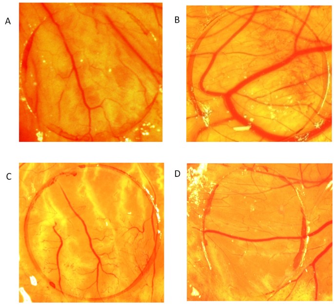
Effect of PHF on the chick chorio-allantoic membrane observed on day 12 of incubation. Photographs showing blood vessels on the Chorio-Allantoic Membrane (CAM) for different groups. A. Normal. B. Treated with VEGF 50 ng. C. Treated with Thalidomide 10 μg along with VEGF 50 ng. D. Treated with 50 μl of PHF along with VEGF 50 ng.
Full size image
Representation of vessel area per coverslip in CAM assay
Full size table
Cautery induced corneal neovascularization
There was no corneal neovascularization observed in the normal (uncauterized) corneas. The PHF group showed significant antiangiogenic activity in the cautery induced corneal neovascularization assay (Figure 2) in comparison to the sham treated group which showed considerable neovascularization in the cornea of rat eyes (Table 2). The positive control (bevacizumab) was observed to significantly inhibit the cautery induced corneal neovascularization at a concentration of 1.25 mg/ml.
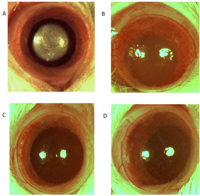
Effect of PHF in cautery induced corneal neovascularization in rats. Photographs showing rat corneas for different groups. A. Cauterized cornea. B. Sham treated cornea. C. Bevacizumab 1.25 mg/ml treated cornea. D. PHF 50 μl treated cornea.
Full size image
Vessel area in the cauterized corneas of different groups
Full size table
Cytotoxicity
The LD50 for taxol was found to be 5.05 μg/ml whereas the PHF did not exhibit any cytotoxicity against the HeLa cancer cell lines.
Hydrocortisone induced chick cataract
The normal (untreated by hydrocortisone) group evinced no development of cataract and control group was graded for stage V (nuclear opacity). In this model PHF showed only a slight protection against cataract as compared to the control group and was graded for stage IV cataract (Figure 3).
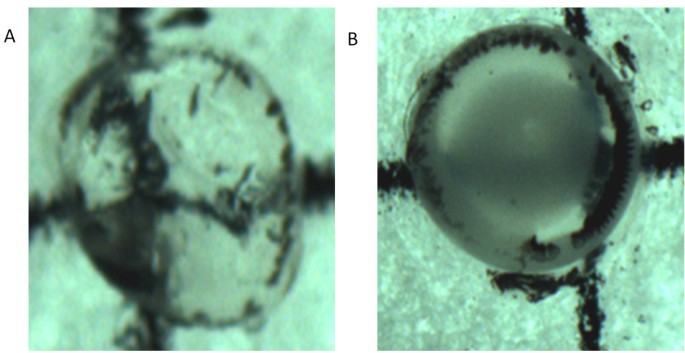
The effect of PHF on lenses in the hydrocortisone induced cataract model in chicks. Photographs showing lenses in different stages of cataract. A: Stage 1: Normal group B: Stage 4: PHF treated group.
Full size image
In vivo selenite induced oxidative model of cataract
The PHF treated group showed significant protection as compared to the control group. In the control group, 100% eyes showed cataract development and were graded for stage IV. Whereas, the eyes treated with PHF showed noticeable delay in the progression of cataract and were graded for stage II. The observations clearly demonstrated the potential of the PHF in delaying the progression of cataract in comparison to the control group.
In vivo galactose induced cataract in rats
At the end of 4 weeks (after galactose feeding) the control group showed significant development of cataract. The lenses in control group depicted stages V respectively, as per Sipple’s gradings. The PHF treated lenses showed slight delay in the development of cataract and were graded for stage III (Figure 4).
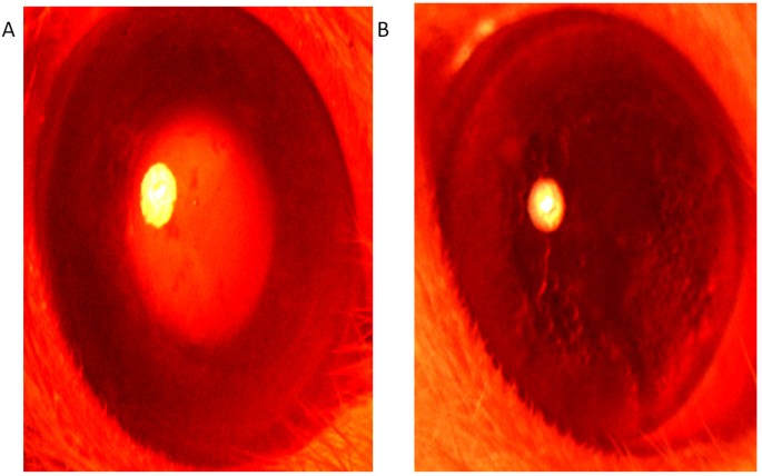
The effect of polyherbal formulation on lenses in the galactose induced cataract model in rats. Photographs showing lenses in different stages of cataract: A: Stage 5: Control group B: Stage 3: PHF group.
Full size image
DPPH radical scavenging assay
The IC50 of the standard (sodium ascorbate) was found to be 5.9 μg/ml. comparatively, the PHF did not show any significant antioxidant activity and the percentage radical scavenging activity up to the maximum of 1.25 ml of PHF was found to be only 26.6%.
Carrageenan induced paw edema
Diclofenac showed maximum inhibition of paw edema (63.6%) after 3 h of its induction via carrageenan. Comparatively, the PHF showed maximum inhibition of paw edema (51.8%) after 3 h (Table 3).
Effect of PHF and diclofenac on carrageenan induced paw edema in rats (after 3 hr)
Full size table
In vitro LTB4 inhibition assay
In the in vitro method involving LTB4 inhibition from human WBC, zileuton was used as a positive control that showed 100% inhibition of LTB4 formation at the tested concentration of 20 μg as compared to untreated control. Among various volumes tried, test group received 20 μl of PHF showed 40% inhibition of LTB4 formation as compared to the control group (Figure 5).
LC-MS/MS chromatograms showing levels of LTB
4
in human WBC’s. A: Control group, B: PHF treated.
Full size image
Intraocular penetration of PHF
PHF tested by IDA protocol showed presence of ar- turmerone, camphor, casticin, curcumin-I, curcumin-II, iso-orientin, luteolin, menthol, quercetin, rosmarinic acid, thymol, wedelolactone, sabinene, γ-terpinene, β- pinene, dipentene. These compounds were further confirmed by MRM.
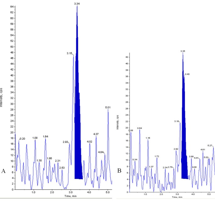
Aqueous humor subjected for optimized MRM for PHF showed presence of camphor, casticin, curcumin-II, quercetin, rosmarinic acid, γ-terpinene, β-pinene and dipentene (Figure 6).

LC-MS/MS chromatogram of aqueous humor sample showing intraocular penetration of PHF in rabbit eyes. MRM chromatogram of compounds with high intensity peaks: (1) Quercetin, (2) γ- terpinene, (3) Rosmarinic acid, (4) β-pinene, (5) Dipentene, (6) Curcumin II.
Full size image
Discussion
The polyherbal eye drop (Itone™) is a sterile topical formulation that has been found to have pH 5.1 and is hypo-osmolar in nature. It has been claimed to protect eyes from continuous strains, glares of light and various forms of pollution upon its instillation. It has also been indicated as an adjunct in various ophthalmic conditions like conjunctivitis, trachoma, blepharitis, keratitis, corneal ulcers, lentricular opacity, myopia, hypermetropia and dacrocystitis [11]. Experiments so far evaluated using PHF showed several properties viz., lack of any acute, subacute CNS toxicity after oral administration in animals, lack of ocular irritancy in rabbits and prominent antimicrobial activity against Staphylococcus aureus and Klebsiella pneumoniae[11]. In clinical studies, the PHF under study has been found to be effective in trachoma, chronic conjunctivitis [12], subjective improvement in refractory errors [13] healing capacity in allergic conjunctivitis [13], viral conjunctivitis [14, 15] and computer vision syndrome [2]. Although, enough literature is available regarding the above therapeutic uses for the PHF, no systematic study is available to substantiate the claim using controlled experiments. Therefore, the present study was conducted to evaluate antiangiogenic, anticataract, anti-inflammatory, antioxidant and cytotoxic potential of the PHF using several experimental models. As PHF under study was also reported to provide relief from iridocyclitis, early senile lenticular opacity and IOP lowering effect in glaucoma, its intraocular penetration was felt as necessary. Therefore, this study was extended to evaluate the intraocular penetration in the rabbit eyes upon topical application using liquid chromatography coupled tandem mass spectrometry.
Ocular angiogenesis is responsible for the majority of irreversible blindness in the developed world. This debilitating complication affects all age groups and characterizes such diverse and widespread diseases as trachoma, retinopathy of prematurity, diabetic retinopathy, neovascular glaucoma and age-related macular degeneration [16]. Angiogenesis is a tightly regulated process involving the development of new blood vessels from pre-existing blood vessels. During development and normal physiological processes such as wound healing and the menstrual cycle, angiogenesis is regulated by endogenous activators and inhibitors [17, 18]. In pathological settings, such as age-related macular degeneration, rheumatoid arthritis, diabetic retinopathy and tumor growth and metastasis, angiogenesis is critical for disease progression [17, 19].
The present study evaluated the antiangiogenic potential of a PHF using the in ovo chick CAM assay – an assay that is capable of evaluating the action of test substance (plant extracts) on angiogenesis [20]. For this assay, VEGF 50 ng was used as an angiogenesis stimulator and was found to significantly induce proliferation of new blood vessels as compared to the normal group. In this assay, PHF was found to have significant antiangiogenic potential at the studied concentration thereby inhibiting VEGF induced proliferation of new blood vessels.
In order to understand the in vivo significane of the above finding an animal study was carried out using chemical cautery induced corneal neovascularization in rats. Neovascularization has been reported as an important pathologic event during the corneal wound healing process [21] and VEGF has been reported to play an important role in its pathogenesis [22]. Neovascularization of cornea may cause loss of corneal transperancy and thereby leads to loss of vision [23]. In the present study, topical instillation of the PHF was found to significantly inhibit chemical cautery induced corneal neovascularization as compared to sham treated control. In this assay, bevacizumab, a known anti-VEGF monoclonal antibody was used as a positive control that showed 67% inhibition of the cautery induced corneal neovascularization in comparison to the sham treated group that showed noticeable corneal neovascularization in the cauterized eyes. The antiangiogenic activity observed for PHF was found to be only 18%, thereby concluding that the antiangiogenic activity observed with PHF is mild in comparison to the potential antiangiogenic compounds like bevacizumab.
Interestingly, the present study revealed that the PHF was found to be safe upon application to the chick embryos and there was no toxicity found. Chorio-allantoic membrane assay is also a well recognized method to study ocular toxicity of drugs [24]. It is assumed as acute irritating effects on the small blood vessels and protein membrane are similar to effect induced by same chemical in the eye [25]. Further to the above observation, lack of any cytotoxicity of the studied PHF was confirmed in HeLa cell lines using MTT assay. In this cytotoxicity assay paclitaxel was used as positive control. This study further confirms lack of any ocular toxicity oberved in majority of the above studies [11].
Inflammatory stimulus is a factor responsible for micro-vascular gowth (neovascularization) during the active pro-angiogenic phase. Therefore, capillary regression is of interest from a clinical perspective [26]. Therapeutic strateties for reducing the vacular growth has been well accepted as a therapeutic option for pathologic angiogenesis in tumors, eye diseases, and inflammation. Anti-inflammatory compounds like COX-2 inhibitors and steroids are reported to have anti-angiogenic potential [27, 28]. Therefore, the present study was extended to carry out anti-inflammatory property of the PHF in the carageenan induced paw edema model of inflammation.
Inflammation is a normal protective responce to tissue injury caused by physical trauma, noxious chemical or microbial agents and involves release of various inflammatory mediators such as leukotrienes and prostaglandins [29–31]. In the present study, anti-inflammatory activity of PHF was tested using carrageenan induced paw edema assay which has been used since time as a standard technique for the screening of anti-inflammatory activity of several herbal extracts [32, 33]. PHF was observed to possess noticeable anti-inflammatory activity, though the activity was comparatively lesser than the positive control (diclofenac). Furthermore, PHF was evaluated for its inhibitory effect on the formation of LTB4 in human WBCs. LTB4 is a known potent inflammatory mediator and has been implicated as a probable cause of chronic ocular inflammation and retinopathy in diabetes [34] and has also been held responsible for the development and progression of experimental autoimmune uveitis [35]. Our study showed that PHF inhibited the formation of LTB4 in human WBCs while the inhibitory percentage was relatively lower in comparison to zileuton which is a known lipoxygenase inhibitor.
The widespread prevalence of diabetes in developing countries is expected to increase the magnitude of blindness due to cataract and the cellular and molecular mechanisms underlying the pathogenesis of cataract showed the involvement of polyol pathway, advanced glycation end products and oxidative stress [36]. The therapeutic agents that are capable of altering the above events are expected to prevent or delay the progression of cataract in order to exhibit the desired anti-cataract activity e.g. chyavanprash [37]. Considering the major pathways involved in the etiology of cataract such as polyol pathway or sorbitol accumulation pathway, anticataract potential of PHF was evaluated using various pharmacological screening models mimicking the major pathways in cataract. In the developing chick embryo model of cataract, glucocorticoid (hydrocortisone) produced higher incidence (>90%) cataractous changes in lenses within 48 h [38]. Cataract formation is caused by oxidative stresses, probably derived from hydrocortisone effects on the main target organ, the liver and can be prevented by radical scavengers including ascorbic acid and insulin. In this model of hyperglycemia and oxidative changes in lens including glutathione depletion are emphasized as factors for the loss of transparency. The present study observed a delay in the progression of cataract in all three models of cataract as compared to their untreated controls. However, the PHF did not show any noticeable free radical scavenging activity in the DPPH assay thereby demarking the presence of any antioxidant potential at the studied concentration. From this observation, it is evident that the mild anticataract activity observed with PHF could be beyond its antioxidant property or as a cumulative effect of antioxidant compounds after repeated administration.
In order to evaluate the possible intraocular penetration of PHF, a study was conducted by taking the aqueous humor after multiple topical instillations for availability of compounds present in PHF. Information dependent acquision method was develpoed and used for the testing of the availability of compounds in PHF. Furthermore presence of compounds in PHF was confirmed by MRM and used for aqueous humor analysis. This study proved that some of the compounds found in PHF have the propansity to cross the cornea to produce the intraocular effects. Among the penetrated compounds curcumin, rosmarinic acid and quercetin are capable of having anticataract activity as reported previously [39–41] whereby curcumin has also been reported for its antiangiogenic potential [42]. However, further studies with isolated and purified compounds are essential to reveal the extent of their penetration for their correlation with observed activity. As the PHF is a proprietary Ayurvedic formulation having many components with very low concentrations, the mild but significant protective effects observed on inflammation, angiogenesis and cataract can have cumulative and synergestic effects while on repeated topical applications in patients for their obvious therapeutic effect with low or no toxicity as observed in clinical studies.
Conclusions
The PHF showed significant antiangiogenic and anti-inflammatory activities and noticeable anticataract activity. However, the PHF did not exhibit any antioxidant potenial and did not show any cytotoxicity against HeLa cancer cell lines. Moreover, the intraocular penetration studies revealed that some of the components of PHF were capable of having transcorneal penetration. The significant antiangiogenic and anti-inflammatory activities evinced by the PHF merits further investigation of PHF for ocular neovascular and inflammatory diseases in humans.
References
-
Indian Council of Medical Research: Ethical guidelines for biomedical research on human participants. 2006, New Delhi, India: Director-General, Indian Council of Medical Research, 50-55.
Google Scholar
-
Biswas NR, Nainiwal SK, Das GK, Langan U, Dadeya SC, Mongre PK, Ravi AK, Baidya P: Comparative randomised controlled clinical trial of a herbal eye drop with artificial tear and placebo in computer vision syndrome. J Indian Med Assoc. 2003, 101 (3): 208-209. 212
CAS
PubMedGoogle Scholar
-
Chatterjee PK, Bairagi D, Roy S, Majumder NK, Paul R, Bagchi S: Comparative randomised active drug controlled clinical trial of a herbal eye drop in computer vision syndrome. J Indian Med Assoc. 2005, 103 (7): 397-398.
PubMed
Google Scholar
-
Ribatti D, Gualandris A, Bastaki M, Vacca A, Iurlaro M, Roncali L, Presta M: New model for the study of angiogenesis and antiangiogenesis in the chick embryo chorioallantoic membrane: the gelatin sponge/chorioallantoic membrane assay. J Vasc Res. 1997, 34 (6): 455-463. 10.1159/000159256.
Article
CAS
PubMedGoogle Scholar
-
Mahoney JM, Waterbury LD: Drug effects on the neovascularization response to silver nitrate cauterization of the rat cornea. Curr Eye Res. 1985, 4 (5): 531-535. 10.3109/02713688508999984.
Article
CAS
PubMedGoogle Scholar
-
Velpandian T, Nirmal J, Gupta P, Vijayakumar AR, Ghose S: Evaluation of calcium dobesilate for its anti-cataract potential in experimental animal models. Methods Find Exp Clin Pharmacol. 2010, 32 (3): 171-179. 10.1358/mf.2010.32.3.1423888.
Article
CAS
PubMedGoogle Scholar
-
Nishigori H, Lee JW, Lwatsuru M: An animal model for cataract research: cataract formation in developing chick embryo by glucocorticoid. Exp Eye Res. 1982, 36: 617-622.
Article
Google Scholar
-
Ostadalova I, Babicky A, Obenberger J: Cataract induced by administration of a single dose of sodium selenite to suckling rats. Experientia. 1978, 34 (2): 222-223. 10.1007/BF01944690.
Article
CAS
PubMedGoogle Scholar
-
Sippel TO: Changes in the water, protein, and glutathione contents of the lens in the course of galactose cataract development in rats. Invest Ophthalmol. 1966, 5 (6): 568-575.
CAS
PubMedGoogle Scholar
-
Blois MS: Antioxidant determinations by the use of a stable free radical. Nature. 1958, 181: 1199-1200. 10.1038/1811199a0.
Article
CASGoogle Scholar
-
ITONE EYE DROPS: Deys. Kolkata, India: Marketing Division, Dey’s Medical Stores (Mfg.) Ltd
-
Das GK, Pandey RM, Biswas NR: Comparative double masked randomised placebo controlled clinical trial of a herbal eye drop preparation in trachoma and conjunctivitis. J Indian Med Assoc. 1995, 93 (10): 383-384.
CAS
PubMedGoogle Scholar
-
Biswas NR, Beri S, Das GK, Mongre PK: Comparative double blind multicentric randomised placebo controlled clinical trial of a herbal preparation of eye drops in some ocular ailments. J Indian Med Assoc. 1996, 94 (3): 101-102.
CAS
PubMedGoogle Scholar
-
Paul AK, Madan S, Gupta V: Clinical evaluation of an indigenous herbal eye drops preparation (Part I). Ind J Clin Prac. 1992, 11: 58-60.
Google Scholar
-
Raizada JK, Mishra L: An extended clinical study with a herbal eye-drop preparation. Ind Med J. 1990, 84: 290-292.
Google Scholar
-
Adamis AP, Aiello LP, D’Amato RA: Angiogenesis and ophthalmic disease. Angiogenesis. 1999, 3 (1): 9-14. 10.1023/A:1009071601454.
Article
CAS
PubMedGoogle Scholar
-
Ferrara N: Vascular endothelial growth factor: basic science and clinical progress. Endocr Rev. 2004, 25 (4): 581-611. 10.1210/er.2003-0027.
Article
CAS
PubMedGoogle Scholar
-
Roskoski R: Vascular endothelial growth factor (VEGF) signaling in tumor progression. Crit Rev Oncol Hematol. 2007, 62 (3): 179-213. 10.1016/j.critrevonc.2007.01.006.
Article
PubMedGoogle Scholar
-
Hoeben A, Landuyt B, Highley MS, Wildiers H, Van Oosterom AT, De Bruijn EA: Vascular endothelial growth factor and angiogenesis. Pharmacol Rev. 2004, 56 (4): 549-580. 10.1124/pr.56.4.3.
Article
CAS
PubMedGoogle Scholar
-
Mathur R, Gupta SK, Singh N, Mathur S, Kochupillai V, Velpandian T: Evaluation of the effect of Withania somnifera root extracts on cell cycle and angiogenesis. J Ethnopharmacol. 2006, 105 (3): 336-341. 10.1016/j.jep.2005.11.020.
Article
PubMedGoogle Scholar
-
Kuo IC: Corneal wound healing. Curr Opin Ophthalmol. 2004, 15 (4): 311-315. 10.1097/00055735-200408000-00006.
Article
PubMedGoogle Scholar
-
Miller JW: Vascular endothelial growth factor and ocular neovascularization. Am J Pathol. 1997, 151 (1): 13-23.
CAS
PubMed
PubMed CentralGoogle Scholar
-
Chang JH, Gabison EE, Kato T, Azar DT: Corneal neovascularization. Curr Opin Ophthalmol. 2001, 12 (4): 242-249. 10.1097/00055735-200108000-00002.
Article
CAS
PubMedGoogle Scholar
-
Luepke NP: Hen’s egg chorioallantoic membrane test for irritation potential. Food Chem Toxicol. 1985, 23 (2): 287-291. 10.1016/0278-6915(85)90030-4.
Article
CAS
PubMedGoogle Scholar
-
Spielmann H: Ocular irritation. In vitro methods in pharmaceutical research. Edited by: Castell JV, Gomez-Lechon MJ. 1997, London: Academic Press, 265-287.
Google Scholar
-
Peebo BB, Fagerholm P, Traneus-Rockert C, Lagali N: Cellular level characterization of capillary regression in inflammatory angiogenesis using an in vivo corneal model. Angiogenesis. 2011, 14 (3): 393-405. 10.1007/s10456-011-9223-3.
Article
CAS
PubMedGoogle Scholar
-
Klenke FM, Gebhard MM, Ewerbeck V, Abdollahi A, Huber PE, Sckell A: The selective Cox-2 inhibitor Celecoxib suppresses angiogenesis and growth of secondary bone tumors: an intravital microscopy study in mice. BMC Cancer. 2006, 6: 9-10.1186/1471-2407-6-9.
Article
PubMed
PubMed CentralGoogle Scholar
-
Oliver A, Ciulla TA: Corticosteroids as antiangiogenic agents. Ophthalmol Clin North Am. 2006, 19 (3): 345-351. v
PubMed
Google Scholar
-
Ohuchi K, Sato H, Tsurufuji S: The content of prostaglandin E and prostaglandin F2alpha in the exudate of carrageenin granuloma of rats. Biochim Biophys Acta. 1976, 424 (3): 439-448. 10.1016/0005-2760(76)90033-3.
Article
CAS
PubMedGoogle Scholar
-
Baccaglini PI, Hogan PG: Some rat sensory neurons in culture express characteristics of differentiated pain sensory cells. Proc Natl Acad Sci U S A. 1983, 80 (2): 594-598. 10.1073/pnas.80.2.594.
Article
CAS
PubMed
PubMed CentralGoogle Scholar
-
Bhitre MJ, Fulmali S, Kataria M, Anwikar S, Kadri H: Anti-inflammatory activity of the fruits of piper longum Linn. Asian J Chemistry. 2008, 20 (6): 4357-4360.
CAS
Google Scholar
-
Winter CA, Risley EA, Nuss GW: Carrageenin-induced edema in hind paw of the rat as an assay for antiiflammatory drugs. Proc Soc Exp Biol Med. 1962, 111: 544-547.
Article
CAS
PubMedGoogle Scholar
-
Jain NK, Singh A, Kulkarni SK: Analgesic, anti-inflammatory and ulcerogenic activity of a zinc-naproxen complex in mice and rats. Pharm Pharmacol Commun. 1995, 5: 599-602.
Article
Google Scholar
-
Talahalli R, Zarini S, Sheibani N, Murphy RC, Gubitosi-Klug RA: Increased synthesis of leukotrienes in the mouse model of diabetic retinopathy. Invest Ophthalmol Vis Sci. 2010, 51 (3): 1699-1708. 10.1167/iovs.09-3557.
Article
PubMed
PubMed CentralGoogle Scholar
-
Liao T, Ke Y, Shao WH, Haribabu B, Kaplan HJ, Sun D, Shao H: Blockade of the interaction of leukotriene b4 with its receptor prevents development of autoimmune uveitis. Invest Ophthalmol Vis Sci. 2006, 47 (4): 1543-1549. 10.1167/iovs.05-1238.
Article
PubMedGoogle Scholar
-
Gupta SK, Selvan VK, Agrawal SS, Saxena R: Advances in pharmacological strategies for the prevention of cataract development. Indian J Ophthalmol. 2009, 57 (3): 175-183. 10.4103/0301-4738.49390.
Article
CAS
PubMed
PubMed CentralGoogle Scholar
-
Velpandian T, Mathur P, Sengupta S, Gupta SK: Preventive effect of Chyavanprash against steroid induced cataract in the developing chick embryos. Phytother Res. 1998, 12: 320-323. 10.1002/(SICI)1099-1573(199808)12:5<320::AID-PTR297>3.0.CO;2-O.
Article
Google Scholar
-
Nishigori H: [Steroid (glucocorticoid)-induced cataract]. Yakugaku Zasshi. 2006, 126 (10): 869-884. 10.1248/yakushi.126.869.
Article
CAS
PubMedGoogle Scholar
-
Suryanarayana P, Krishnaswamy K, Reddy GB: Effect of curcumin on galactose-induced cataractogenesis in rats. Mol Vis. 2003, 9: 223-230.
CAS
PubMedGoogle Scholar
-
Mohan M, Gupta SK, Agnihotri S, Joshi S, Uppal RK: Anticataract action of topical quercetin and myricetin in galactosemic rats. Med Sci Res. 1988, 16: 685-686.
CAS
Google Scholar
-
Park SU, Uddin MR, Xu H, Kim YK, Lee SY: Biotechnological applications for rosmarinic acid production in plant. African J Biotech. 2008, 7: 4959-4965.
CAS
Google Scholar
-
Arbiser JL, Klauber N, Rohan R, van Leeuwen R, Huang MT, Fisher C, Flynn E, Byers HR: Curcumin is an in vivo inhibitor of angiogenesis. Mol Med. 1998, 4 (6): 376-383.
CAS
PubMed
PubMed CentralGoogle Scholar
Pre-publication history
-
The pre-publication history for this paper can be accessed here:http://www.biomedcentral.com/1472-6882/13/1/prepub
Download references
Acknowledgement
We thank Dey’s Medical Store, Kolkata for providing preservative free Itone™ and academic research grant for conducting this scientific study.
Author information
Authors and Affiliations
-
Department of Ocular Pharmacology and Pharmacy, Dr. Rajendra Prasad Centre for Ophthalmic Sciences, All India Institute of Medical Sciences, Room No: 634, 6th floor, New Delhi, 110029, India
Thirumurthy Velpandian, Pankaj Gupta, Alok Kumar Ravi & Hanuman Prasad Sharma
-
Department of Pharmacology, All India Institute of Medical Sciences, New Delhi, 110029, India
Nihar Ranjan Biswas
Authors
- Thirumurthy Velpandian
You can also search for this author in
PubMed Google Scholar - Pankaj Gupta
You can also search for this author in
PubMed Google Scholar - Alok Kumar Ravi
You can also search for this author in
PubMed Google Scholar - Hanuman Prasad Sharma
You can also search for this author in
PubMed Google Scholar - Nihar Ranjan Biswas
You can also search for this author in
PubMed Google Scholar
Corresponding author
Correspondence to
Thirumurthy Velpandian.
Additional information
Competing interests
Dey’s Medical Store, Kolkata is the manufacturer of Itone™ who has provided preservative free formulation and academic research grant for conducting this scientific study. The authors hereby declare that the manufacturer had no role or interest in the design, conduct and interpretation of the study.
Authors’ contributions
VT designed the experiments and drafted the manuscript. GP carried out the pharmacological screening of PHF. RAK carried out the anticataract screening of PHF. SHP carried out intraocular penetration of PHF and anti-inflammatory screening using carrageenan induced paw edema assay. BNR participated in the design and co-ordination of the study. All the authors read and approved the final manuscript.
Authors’ original submitted files for images
Rights and permissions
This article is published under license to BioMed Central Ltd. This is an Open Access article distributed under the terms of the Creative Commons Attribution License (http://creativecommons.org/licenses/by/2.0), which permits unrestricted use, distribution, and reproduction in any medium, provided the original work is properly cited.
Reprints and Permissions
About this article
Cite this article
Velpandian, T., Gupta, P., Ravi, A.K. et al. Evaluation of pharmacological activities and assessment of intraocular penetration of an ayurvedic polyherbal eye drop (Itone™) in experimental models.
BMC Complement Altern Med 13, 1 (2013). https://doi.org/10.1186/1472-6882-13-1
Download citation
-
Received: 04 August 2012
-
Accepted: 20 December 2012
-
Published: 02 January 2013
-
DOI: https://doi.org/10.1186/1472-6882-13-1
Keywords
- Polyherbal
- Antiangiogenic
- Anti-inflammatory
- Anticataract
- Antioxidant
- Cytotoxicity
- LC-MS/MS